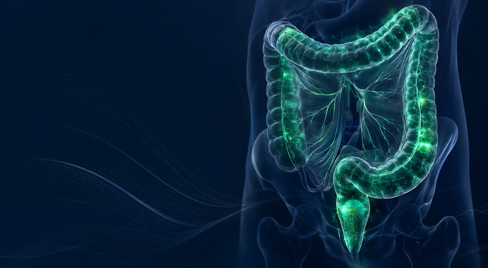
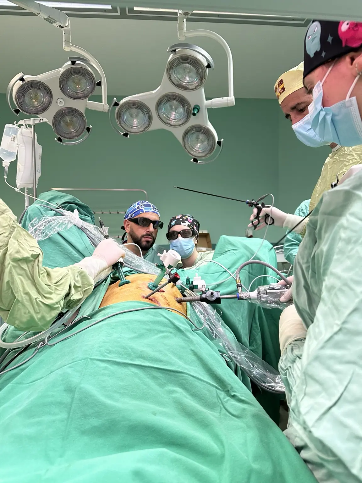
Treatment is planned for the individual. Tumour location and stage, imaging findings, overall health and the patient's priorities all shape the operation. The aim is a safe oncological resection while limiting treatment burden where possible and protecting bowel function and quality of life.
Colon or rectal cancer may cause no symptoms at an early stage. A persistent, unexplained change in bowel function deserves medical assessment, although it does not necessarily mean cancer.
01
CHANGE IN BOWEL HABIT
New persistent diarrhoea, constipation or a change in bowel frequency.
02
BLOOD OR BLEEDING
Blood in the stool or bleeding from the rectum should be assessed.
03
INCOMPLETE EMPTYING
A feeling that the bowel has not emptied or a constant urge to pass stool.
04
ABDOMINAL SYMPTOMS
Persistent pain, cramps, pressure or bloating without a clear explanation.
05
WEIGHT LOSS
Unexplained weight loss or reduced appetite.
06
POSSIBLE ANAEMIA
Fatigue, breathlessness or dizziness may reflect blood loss and anaemia.
These symptoms can have many benign causes. Diagnosis requires clinical assessment and, when indicated, endoscopy or imaging. Screening remains important even when there are no symptoms.
01 · FROM DIAGNOSIS TO PLAN
One oncology plan, designed for one patient
Treatment is organised step by step. In rectal cancer, high-quality MRI and multidisciplinary review come before any definitive decision.
01PRECISE STAGING
Colonoscopy and biopsy, CT of chest–abdomen–pelvis, CEA and dedicated pelvic MRI for rectal cancer. Molecular or genetic testing when indicated.
02MULTIDISCIPLINARY REVIEW
Surgeon, radiologist, pathologist, medical oncologist and radiation oncologist build one shared strategy rather than isolated decisions.
03TAILORED OPERATION
Resection extent, laparoscopic or open access and reconstruction are selected according to location, stage and individual anatomy.
04PATHOLOGY & FOLLOW-UP
Structured specimen assessment, review of final stage and an organised plan for adjuvant treatment or surveillance.
02 · COLON CANCER
A different colectomy for each location
There is no single operation for the entire colon. Resection follows the vascular and lymphatic anatomy of the segment that contains the tumour.
CME
Oncological colectomy aims for an intact mesocolic-plane resection with adequate regional lymphadenectomy. Formal Complete Mesocolic Excision (CME) with central vascular ligation is not automatic for every patient; it is selected according to tumour location, vascular anatomy and the experience of the surgical team.
Anterior view · R / L: patient's sides
Illustrative resection
Bowel preserved
Illustrative lesion
Division points
Anastomosis
Schematic illustrations to explain surgery. The extent of resection, reconstruction and possible stoma are individualised. Vessels, mesocolon and lymph-node clearance boundaries are not shown.
01Caecum · Ascending colon
01What is removed
02How the bowel is joined
Illustrative reconstructionIleocolic anastomosis
RIGHT HEMICOLECTOMY
Removal of the right colon and its lymphatic field, followed by an ileocolic anastomosis.
02Hepatic flexure · Proximal transverse
01What is removed
02How the bowel is joined
Illustrative reconstructionIleocolic anastomosis
EXTENDED RIGHT HEMICOLECTOMY
A wider oncological resection when tumour position requires coverage of adjacent vascular and nodal territories.
03Mid transverse colon
01What is removed
02How the bowel is joined
Illustrative reconstructionColocolic anastomosis
SEGMENTAL TRANSVERSE COLECTOMY
Selected segmental resection with careful planning of perfusion, margins and lymphadenectomy.
04Splenic flexure · Descending colon
01What is removed
02How the bowel is joined
Illustrative reconstructionColocolic anastomosis
LEFT HEMICOLECTOMY
Oncological removal of the left colon with reconstruction adapted to bowel length and perfusion.
05Sigmoid colon
01What is removed
02How the bowel is joined
Illustrative reconstructionColorectal anastomosis
SIGMOID COLECTOMY
Laparoscopic oncological sigmoid resection with reconnection of healthy colon to the rectum.
Removal of most of the colon while preserving a selected distal segment when disease distribution, oncological safety and perfusion permit. Reconstruction is individualised.
07Diffuse colonic disease · selected indications
01What is removed
02How the bowel is joined
Illustrative reconstructionIleorectal anastomosis
TOTAL COLECTOMY
Removal of the entire colon with preservation of the rectum and an ileorectal anastomosis when oncologically and functionally appropriate. A different reconstruction or a stoma may be required in other circumstances.
Illustrative reconstructionTransverse colon to rectum
SPLENIC FLEXURE & COMPLEX RECONSTRUCTION
Resection extent and reconstruction — including a transverse-to-rectal anastomosis or modified Deloyers strategy — are selected according to disease distribution, vascular anatomy, perfusion and the ability to create a tension-free anastomosis.
03 · ONCOLOGICAL PLANES
CME for the colon. TME for the rectum.
Both principles share one philosophy: remove the tumour together with its natural lymphovascular envelope along clean anatomical planes.
CME · COLON
COMPLETE MESOCOLIC EXCISION
Oncological colectomy follows the embryological mesocolic plane with adequate regional lymphadenectomy. Formal CME and central vascular ligation are used when appropriate for tumour location, anatomy and the individual case.
Intact mesocolic envelope
Central vascular approach where appropriate
Adequate margins and nodal staging
TME · RECTUM
TOTAL MESORECTAL EXCISION
For mid and low rectal cancer, the rectum is removed with the mesorectum intact through a meticulous, nerve-preserving pelvic dissection.
Clear circumferential margin
Pelvic autonomic nerve preservation
Oncological resection with functional priorities
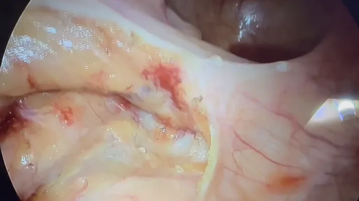
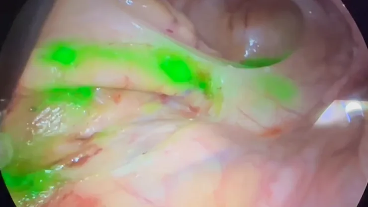
WHITE LIGHT · SAME OPERATIVE FIELDNIR · ICG LYMPHATIC MAPPING
Real intraoperative recording: lymphatic drainage and nodal mapping during sigmoid cancer surgery. A selective adjunct — not a substitute for indicated oncological lymphadenectomy.
04 · FLUORESCENCE-GUIDED SURGERY
ICG fluorescence: real-time information
Indocyanine green (ICG) is visualised with a near-infrared camera. Different routes and protocols may support lesion localisation, selective lymphatic mapping or perfusion assessment. It does not replace oncological resection or surgical judgement.
HOW ICG WORKS — IN PLAIN LANGUAGE
01 · THE DYE
Indocyanine green (ICG) is a fluorescent dye used as an additional imaging tool during surgery.
02 · THE ADMINISTRATION
The route depends on the purpose: intravenous administration for perfusion assessment, or local/peritumoural administration under a dedicated protocol for lymphatic mapping.
03 · THE NIR CAMERA
A near-infrared camera detects the signal and displays it in real time. This is fluorescence, not fluoroscopy, and uses no ionising radiation.
04 · THE INTERPRETATION
The finding is interpreted alongside anatomy and other intraoperative information. Dose, timing, haemodynamic state and camera settings can alter the image.
01
BEFORE SURGERY
Marking & localisation
In selected cases, endoscopic submucosal ICG marking before laparoscopy can support lesion localisation. Fluorescence imaging of liver metastases uses different, dedicated protocols and is not illustrated here.
02
DURING ONCOLOGICAL RESECTION
Selective lymphatic mapping
Depending on the protocol, peritumoural administration may visualise lymphatic drainage. This is a selective adjunct; it does not replace indicated oncological lymphadenectomy or determine resection extent on its own.
03
BEFORE & AFTER ANASTOMOSIS
Perfusion assessment
Intravenous ICG allows assessment of blood flow at both bowel ends before they are joined and, where appropriate, after the anastomosis is complete. The image may support a safer transection point.
05 · RECTAL CANCER
From local excision to complex pelvic surgery
Distance from the sphincter, depth of invasion, MRI findings and response to preoperative treatment determine whether the best strategy is local excision, TME, sphincter-preserving reconstruction or a different pathway.
TME
Sphincter preservation is an important goal, but never at the expense of oncological margins. Each decision follows complete staging, MDT review and an honest discussion about function, the possibility of a temporary or permanent stoma and the alternatives.
Schematic orientation only — not an operative plan for an individual patient. Tumour position, vascular anatomy, resection extent and reconstruction are patient-specific.
01Selected early lesions
TEM / TAMIS · LOCAL EXCISION
Transanal excision for carefully selected low-risk cT1N0 tumours or complex polyps. It provides no nodal staging; adverse histology may require subsequent radical resection.
02Upper rectum
HIGH ANTERIOR RESECTION
Removal of the upper rectum with oncologically adequate partial mesorectal excision (PME) and colorectal anastomosis.
03Mid and low rectum
LOW ANTERIOR RESECTION
TME with reconnection of the colon to the remaining rectum, often protected temporarily with a diverting ileostomy.
04Very low rectum
ULTRA-LOW / COLOANAL ANASTOMOSIS
Reconstruction close to the anal canal when safe oncological margins and acceptable expected function can both be achieved.
05Selected very low tumours
INTERSPHINCTERIC RESECTION
Removal of part of the internal sphincter with coloanal reconstruction only when the oncological findings and baseline function make it appropriate.
06Very low tumour or technically demanding pelvis
TaTME · CECIL TWO-TEAM
TaTME changes the access route, not the required oncological extent of TME. It is reserved for selected patients in specialist centres with structured training and proctoring, institutional governance and outcome audit, because of its learning curve and technique-specific risks.
07When the sphincter cannot be preserved
ABDOMINOPERINEAL RESECTION / ELAPE
Radical removal of the rectum and anus with a permanent colostomy when sphincter-preserving surgery is not oncologically safe.
08Locally advanced or recurrent tumour
EN BLOC MULTIVISCERAL RESECTION
Complex multidisciplinary removal of adjacent organs in selected cases where complete oncological clearance remains a realistic goal. The simplified diagram shows the pelvic resection field; adjacent organs are not depicted.
Protective ileostomy for rectal cancer
In selected patients, a protective loop ileostomy is created after low anterior resection. Stool is diverted into a pouch to reduce the load on the low bowel join while it heals.
Anterior view · R / L: patient's sides
01Before: tumour and illustrative resection
02After: bowel join and ileostomy
1Rectal tumour
2Low colorectal anastomosis
3Protective loop ileostomy
4Collection pouch
Stool flow into the pouch
Two openings in the same ileal loop
AProximal opening: stool outlet
BDistal opening: continues towards the colon
Schematic example after low anterior resection. A protective ileostomy is not needed for every rectal tumour. It is usually intended to be temporary; reversal is considered after healing has been assessed and when it is safe. It does not eliminate the risk of an anastomotic leak.
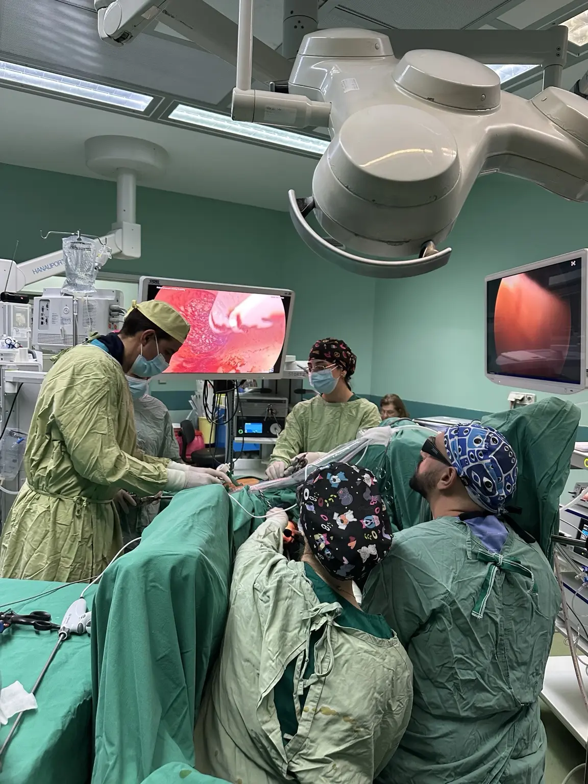
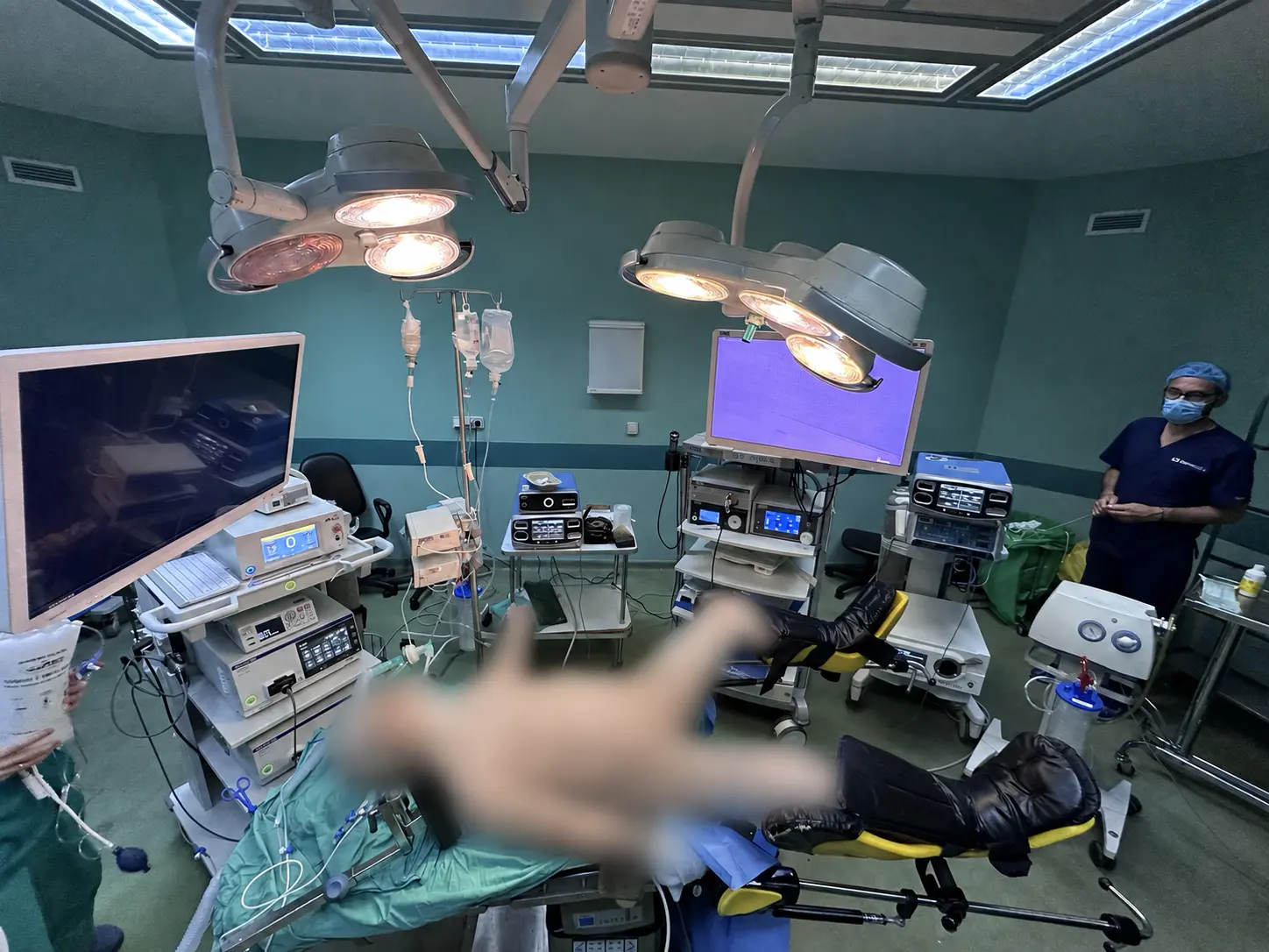
Modern laparoscopic imaging is most effective when theatre roles are clear and the whole team shares one plan before the operation begins.
MINIMALLY INVASIVEAdvanced laparoscopic surgery with high-definition imaging.TWO-TEAM WORKFLOWA coordinated two-team approach for selected, technically demanding rectal operations.PATIENT PRIVACYAn organised high-technology environment with the patient's identity protected.
07 · DECISIONS THAT MATTER
Questions that should be answered before surgery
Technique is only part of the decision. Every patient should understand why a strategy is recommended and what it may mean for life after surgery.
01Will the operation be laparoscopic?
A minimally invasive approach is preferred when technically and oncologically appropriate. Open surgery remains the right option for some very large or locally invasive tumours, for emergency presentations and for anatomically complex cases. Safety and completeness come before incision size.
02Will I need a stoma?
Most planned colectomies do not require a permanent stoma. A very low anastomosis may need a temporary diverting ileostomy. An end colostomy is required after abdominoperineal resection and may be the safest option after a Hartmann procedure or another non-restorative resection, particularly in obstruction, perforation or another emergency, or when frailty and other medical conditions make an anastomosis unsafe. The decision is individualised and, whenever possible, discussed before surgery.
03Can rectal cancer be managed without surgery?
In strictly selected patients with a complete clinical response after total neoadjuvant therapy (TNT), a Watch & Wait strategy may be discussed. This is not passive observation: it requires an expert MDT, rigorous criteria and intensive long-term surveillance.
04How is the exact operation selected?
By tumour location, stage, vascular anatomy, sphincter function, previous surgery or radiotherapy, other medical conditions and the patient's priorities. The same diagnosis can require a different plan in two different people.
EVIDENCE · STANDARDS · TRANSPARENCY
The standards behind this page
The page is structured around current guidance and quality standards from international surgical organisations. External links open the original source material.
This page provides general information and does not replace personalised medical assessment. The indication, extent and technique of each operation are decided after full review of the individual case.
ELIXIS · NEXT STEP
A clear plan starts with the complete picture
Bring your colonoscopy, histology, CT and — for a rectal tumour — pelvic MRI to the appointment. For electronic transfer, contact the practice first so that a secure method can be provided; do not send medical records by ordinary email. The options, benefits and limitations can then be discussed.